Duration 19:17
Uterus Anatomy | Structure & Function of Uterus | What is womb | Female reproductive system | MCQ
Published 5 Aug 2021
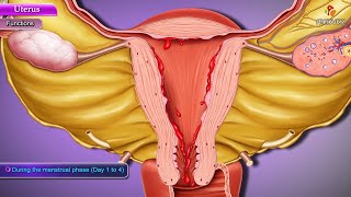
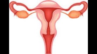
In this lecture I discuss the anatomy and physiology of uterus. As well as question and answer of Uterus also discuss. Uterus The uterus (womb) serves as part of the pathway for sperm deposited in the vagina to reach the uterine tubes. It is also the site of implantation of a fertilized ovum, development of the fetus during pregnancy, and labor. During reproductive cycles when implantation does not occur, the uterus is the source of menstrual flow. Anatomy of the Uterus Situated between the urinary bladder and the rectum, the uterus is the size and shape of an inverted pear . In females who have never been pregnant, it is about 7.5 cm (3 in.) long, 5 cm (2 in.) wide, and 2.5 cm (1 in.) thick. The uterus is larger in females who have recently been pregnant, and smaller (atrophied) when sex hormone levels are low, as occurs after menopause. Anatomical subdivisions of the uterus include: (1) a dome shaped portion superior to the uterine tubes called the fundus, (2) a tapering central portion called the body, and (3) an inferior narrow portion called the cervix that opens into the vagina. Between the body of the uterus and the cervix is the isthmus , a constricted region about 1 cm (0.5 in.) long. The interior of the body of the uterus is called the uterine cavity, and the interior of the cervix is called the cervical canal. The cervical canal opens into the uterine cavity at the internal os (os mouthlike opening) and into the vagina at the external os. Normally, the body of the uterus projects anteriorly and superiorly over the urinary bladder in a position called anteflexion. The cervix projects inferiorly and posteriorly and enters the anterior wall of the vagina at nearly a right angle. Several ligaments that are either extensions of the parietal peritoneum or fibromuscular cords maintain the position of the uterus . The paired broad ligaments are double folds of peritoneum attaching the uterus to either side of the pelvic cavity. The paired uterosacral ligaments, also peritoneal extensions, lie on either side of the rectum and connect the uterus to the sacrum. The cardinal (lateral cervical) ligaments are located inferior to the bases of the broad ligaments and extend from the pelvic wall to the cervix and vagina. The round ligaments are bands of fibrous connective tissue between the layers of the broad ligament; they extend from a point on the uterus just inferior to the uterine tubes to a portion of the labia majora of the external genitalia. Although the ligaments normally maintain the anteflexed position of the uterus, they also allow the uterine body enough movement such that the uterus may become malpositioned. A posterior tilting of the uterus, called retroflexion (retro- backward or behind), is a harmless variation of the normal position of the uterus. There is often no cause for the condition, but it may occur after childbirth. Histology of the Uterus Histologically, the uterus consists of three layers of tissue: perimetrium, myometrium, and endometrium . The outer layer—the perimetrium or serosa—is part of the visceral peritoneum; it is composed of simple squamous epithelium and areolar connective tissue. Laterally, it becomes the broad ligament. Anteriorly, it covers the urinary bladder and forms a shallow pouch, the vesicouterine pouch . Posteriorly, it covers the rectum and forms a deep pouch between the uterus and urinary bladder, the rectouterine pouch or pouch of Douglas— the most inferior point in the pelvic cavity. The middle layer of the uterus, the myometrium (myo- muscle), consists of three layers of smooth muscle fibers that are thickest in the fundus and thinnest in the cervix. The thicker middle layer is circular; the inner and outer layers are longitudinal or oblique. During labor and childbirth, coordinated contractions of the myometrium in response to oxytocin from the posterior pituitary help expel the fetus from the uterus. Various playlist of our Channel Anitmicrobial agent Pharmacology /watch/fJGFQggpct1zmetlCc5qUW3zC7eZljiwLP=tsil&cbOpL_OEOjOEF Human Anatomy & Physiology Lectures Playlist /watch/U1B4E1JDeXmEvp24s3tqzU3zC7eZljiwLP=tsil&UxgSCyl8d3884 FACEBOOK: https://www.facebook.com/vivekjainsir/ INSTAGRAM: https://www.instagram.com/tutorbox3/ You tube Channel Link: /tutorboxchannel .
Category
Show more


Comments - 10
Related videos for Uterus Anatomy | Structure & Function of Uterus | What is womb | Female reproductive system | MCQ: